Craniovertebral Angle: How Forward Head Is Measured and What's Normal
The craniovertebral angle (CVA) is read from a side photo: a horizontal line through the C7 vertebra at the base of the neck, a line from C7 to the ear’s tragus, and the angle between them. A smaller angle means a more forward head. No single value is normal. Studies set the forward-head cut-off anywhere from 44° to 54°, and healthy adults in two recent studies averaged 48.8° seated and 50.0° standing, so age and body position matter.
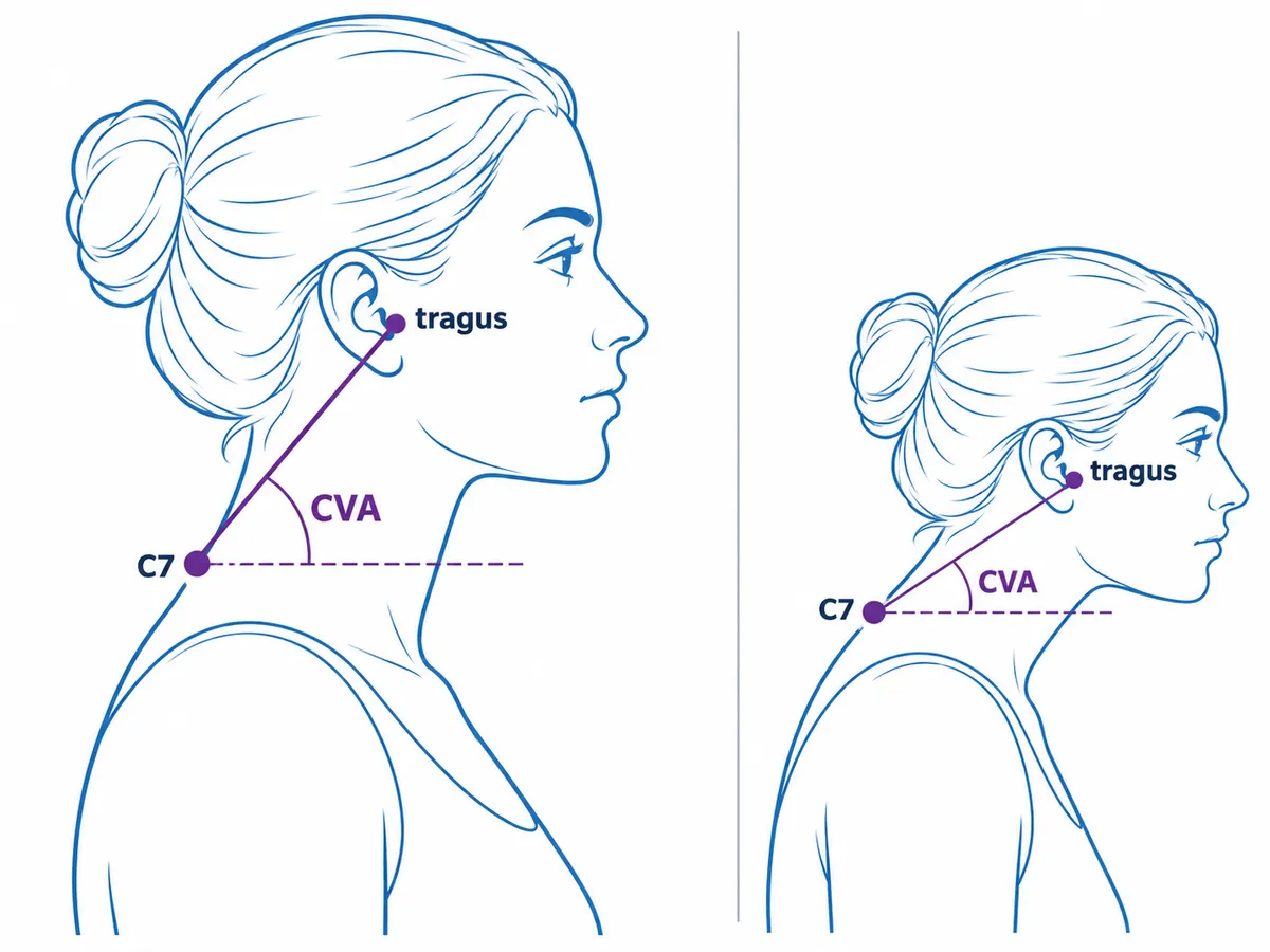
How the craniovertebral angle is measured
In clinics and research, the craniovertebral angle is usually measured by photogrammetry: angles drawn on a marked side photo. Camera setups vary between studies, but the steps are the same.
- Mark C7. A clinician finds the C7 spinous process, the bony bump at the base of the neck, by touch and sticks a small marker on it.
- Mark the tragus. A second marker goes on the tragus, the small flap of cartilage in front of the ear canal, on the side facing the camera.
- Stand relaxed and look ahead. In a 2024 study, Titcomb and colleagues asked people to look straight ahead at a wall and hold still for five seconds before each photo.
- Set the camera level and side-on. Published setups differ: 3 m away at C7 height, standing (Titcomb, 2024); 1.4 m away at shoulder level, standing (Mylonas et al., 2025); 1 m away at shoulder height, seated (Zárate-Tejero et al., 2024).
- Draw and read the angle. In image software such as Kinovea or ImageJ, draw a horizontal line through the C7 marker and a line from C7 to the tragus marker. The angle between them is the CVA.
Done this way, the reading is repeatable. Four physiotherapists measuring 30 university students agreed almost perfectly, with an intraclass correlation (ICC, where 1.0 means identical readings) of 0.98 between examiners, and 0.95 when one examiner repeated the measurement the next day (Mylonas et al., 2025). A 2022 systematic review found the strongest reliability evidence for this photo method among the forward-head measures it covered, but found the evidence on validity inconclusive.
Validity matters because the CVA describes where the head sits over the base of the neck, not the curve of the neck bones. In 120 adults with chronic myofascial pain, all with a CVA of 50° or less, the photo CVA and an X-ray measure of how far the neck sits forward (the C2–C7 sagittal vertical axis) were only loosely related (r = −0.549, which the authors call weak). The authors concluded that the two describe different aspects of neck alignment and that the CVA cannot replace X-ray measures of the cervical curve (Oakley et al., 2024).
What is a normal craniovertebral angle?
No cut-off is agreed. Reviewing the literature, Titcomb and colleagues (2024) found forward head posture defined anywhere from a CVA of 44° or less to 54° or less, and “normal” starting above 53° in one study and at 55° or more in another. The same authors note that a CVA below 50° typically counts as forward head posture.
The trouble with any single cut-off is that healthy people sit right on it. Titcomb’s 98 young adults without known pathology averaged 50.0° standing. In 122 healthy adults aged 18 to 75 photographed seated, recruited as about 10 men and 10 women per age group, Zárate-Tejero and colleagues (2024) found an average of 48.8°, about 2.5° lower in men than in women, and falling by about 1.6° per decade of age. These were seated photos, so they don’t test a standing cut-off directly, but they show how far the healthy average moves with age.
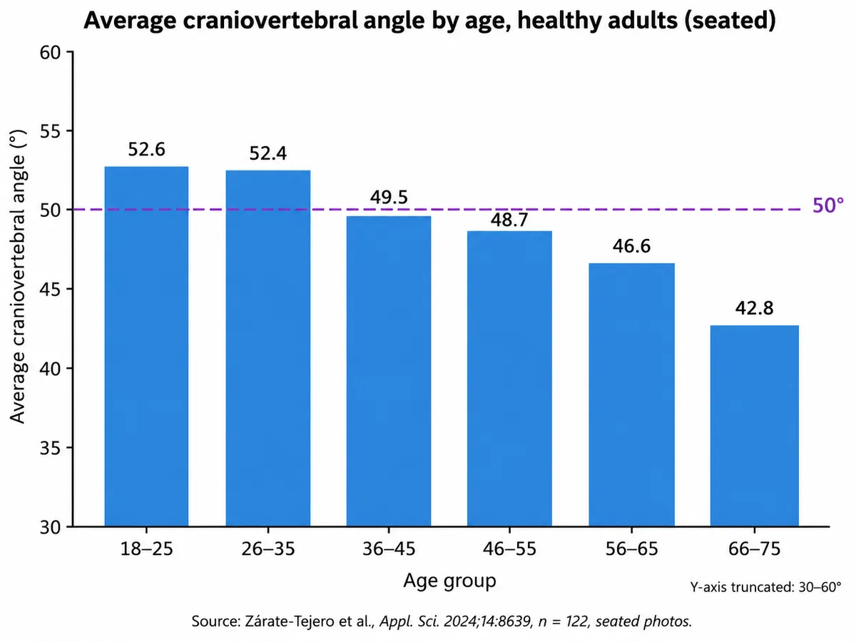
| Age group | Average CVA, seated |
|---|---|
| 18–25 | 52.6° |
| 26–35 | 52.4° |
| 36–45 | 49.5° |
| 46–55 | 48.7° |
| 56–65 | 46.6° |
| 66–75 | 42.8° |
Read against these averages, a seated reading of 45° is close to typical for someone in their 60s but almost 8° below the average for someone in their early 20s. Our reading of these studies: a single CVA between roughly 45° and 55° says little on its own. It means more when it is compared with people of the same age measured the same way, and most when it is compared with the same person’s earlier readings.
Group averages differ with neck pain only in some age groups. In a 2019 meta-analysis, adults aged 18 to 50 with neck pain had a CVA 4.84° smaller on average than adults without it (3 studies, 203 people, with large differences between studies). Adolescents showed no difference, nor did the single study of adults over 50, and pooling all seven CVA studies across ages gave no significant difference (Mahmoud et al., 2019). These are one-time comparisons between groups: they don’t show that a smaller angle causes neck pain, and they give no threshold that tells one person whether their neck will hurt.
Sitting or standing changes the number
Body position moves the craniovertebral angle, but studies disagree on the direction. In Titcomb’s young adults, the average was 2.2° lower seated than standing (47.8° vs 50.0°). A 2016 study of 50 young adults (Shaghayegh Fard et al.) found the opposite, a larger angle when seated, and so did a 2020 study of 70 older adults with an average age of 68 (Abbaszadeh-Amirdehi et al.: 48.1° standing, 52.0° seated).
Both young-adult studies recommend standing as the standard position. Whichever you use, compare a standing reading only with standing cut-offs and other standing readings. The age table above comes from seated photos, so a standing reading isn’t directly comparable with it.
A different angle with the same name
Searching for the craniovertebral angle also turns up values around 150°. Those belong to a different measurement that uses the same name:
| Posture CVAThis article | MRI craniovertebral angleClivus-canal angle | |
|---|---|---|
| Measured on | Side photo (or X-ray) | Side-view MRI |
| Lines | Horizontal through C7; C7 to ear tragus | Along the clivus (skull base); along the back of C2 |
| Typical values | Healthy adults averaged 48.8–50.0° in two studies | Normally 150° in flexion to 180° in extension |
| What it describes | Head position over the base of the neck | Alignment where the skull meets the spine |
The MRI version comes from radiology: a 2014 study of 205 people with cervicogenic headache measured it on MRI, and its control group averaged 153.3° (Çoban et al., 2014). If a report gives a craniovertebral angle near 150°, it is not a forward-head reading and can’t be compared with the 44–54° cut-offs.
How PosturaScreen’s Forward Head metric relates to the craniovertebral angle
PosturaScreen, the product behind this article, cannot measure the craniovertebral angle. Its pose model finds 17 standard body keypoints (nose, eyes, ears, shoulders, elbows, wrists, hips, knees and ankles), and none of them is C7 or the tragus. From a side photo it reports two related metrics instead:
- Forward Head is the angle of the line from the shoulder keypoint to the ear keypoint, measured from vertical. The number has no sign: it grows as the ear moves horizontally away from directly above the shoulder, which in forward head posture means forward. The report range is under 12°, and the metric carries the
approxtag (saved reports label it “Estimated”). - Ear-Shoulder is the horizontal distance between the same two points in centimetres, scaled by assuming a 50 cm shoulder-to-hip torso. The report range is under 2.5 cm.
The 12° line is PosturaScreen’s own screening threshold, not a published CVA value. A note in the analysis code explains it: 2.5 cm of forward offset over a neck of about 12 cm gives about 12° (the arctangent of 2.5 ÷ 12 is 11.8°). There is no formula to turn Forward Head into a CVA either. The shoulder keypoint marks the shoulder, not the spine, and the two angles use different reference lines, so they move in opposite directions: Forward Head grows as the head moves forward, while the CVA shrinks.
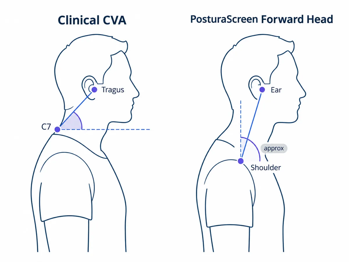
The two PosturaScreen numbers can also disagree with each other. In the sample report, made for a fictional client, Forward Head reads 11.1°, inside its range, while Ear-Shoulder reads 4.0 cm, outside its range. Both come from the same ear and shoulder points. By our calculation (4.0 ÷ tan 11.1°), those two values put the ear about 20 cm above the shoulder point on the report’s scale, well above the roughly 12 cm the 12° threshold assumes. The taller that distance, the smaller the angle a given forward offset makes, so in that report Ear-Shoulder flags and Forward Head doesn’t.
Can an angle from automatically detected points track forward head at all? Indirect evidence says yes, with limits. In 2025, Park and colleagues tested a different AI posture tool, which places C7 and the centre of the head itself, on 36 patients with neck pain or alignment problems, in a small pilot study in which three of the authors work for the tool’s maker. Its forward-head angle correlated with the X-ray CVA at r = −0.71: strongly related, not identical. It tested another algorithm, so it doesn’t validate PosturaScreen’s numbers. The formulas for every metric are on the methodology page, and the posture metrics glossary explains each one. For what forward head posture looks like and how common it is, see the forward head posture guide.
How to track a forward head reading over time
For one person, change over time tells you more than any cut-off. To make two readings comparable:
- Same position. Standing is the position both young-adult studies above recommend. Don’t mix standing and seated readings.
- Same setup. Use the same spot, camera height and distance, with the camera level and square to your side. PosturaScreen checks keypoint confidence and that your whole body is in frame, but it doesn’t detect a tilted camera; the guide to taking posture photos covers the setup.
- Relaxed, eyes ahead, ear visible. Tie back long hair and avoid high collars, and don’t straighten up for the photo.
- Treat small changes with caution. In a 2020 study of a phone app that measures the CVA from marked photos (44 young adults, standing), a change had to exceed about 5° to count as more than measurement error: the minimum detectable change ranged from 4.5° to 5.9° across examiners and sessions. That figure belongs to that app and protocol. It cannot establish a change threshold for PosturaScreen’s Forward Head, which uses different landmarks and has no published minimum detectable change of its own.
A photo angle can’t explain symptoms. The NHS advises seeing a GP if neck pain or stiffness hasn’t gone after a few weeks, or comes with pins and needles or a cold arm; a licensed physiotherapist, physician, or chiropractor can then put a CVA reading in context. If your report flags Forward Head without symptoms, what to do after a posture screen sets out a track-and-recheck approach.
How this article was made: the study figures come from the papers linked in the text, opened in October 2026. The description of PosturaScreen’s metrics comes from its analysis code and the published methodology; the sample report is fictional. It is for posture education, is not medical advice and is not a substitute for a clinical evaluation. PosturaScreen is a screening and tracking tool, not a diagnostic device. See our editorial standards for how articles are written.
Free posture screening, in seconds.
Start with one photo. Add the second for all 17 metrics. No account or app required.
Start a free posture screen